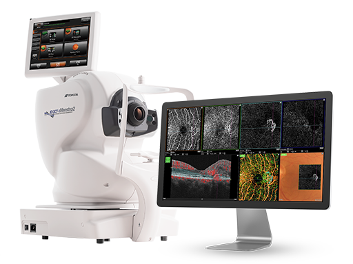
OCT-A is not available at Marion. Call 319-462-4891 to arrange imaging at the appropriate Sigma office.
Retinal Layers.
Blood Vessels.
No Injected Dye.
The retina is the light-sensitive lining at the back of your eye. OCT uses light to show its layers, helping us look for swelling and other changes. OCT-A shows blood flow in its tiny vessels without injecting dye.
For age-related macular degeneration (AMD), which affects the detailed central vision used for reading and recognizing faces, OCT helps us look for damage and fluid, while OCT-A helps reveal abnormal blood vessels associated with wet AMD. Used with your eye exam, these images help distinguish dry changes from signs of wet AMD. They also help us assess damage from diabetes and other retinal conditions.
The OCT-A scan is noninvasive (without cutting or piercing the eye) and takes seconds. Rest your chin, hold still, and look at a target while the scan captures fine detail without injected dye or a bright camera flash. Other parts of your examination may include photographs or dilation (drops that widen your pupils so we can see inside your eyes). Your doctor explains what the findings mean and whether further testing or treatment is needed.
Macular Degeneration & Medical Eye Care